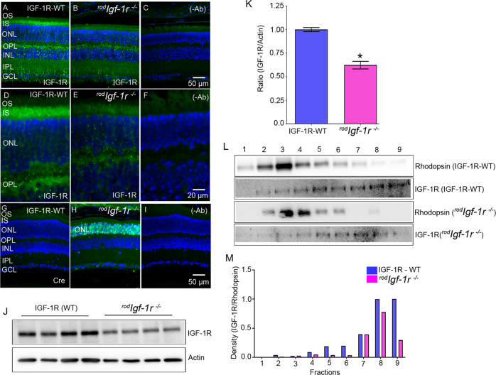
Fig. 1. Expression of IGF-1R in wild-type and rodIgf-1r−/− mice.
Prefer-fixed sections of 2-month-old WT (A, D, G) and rodIgf-1r−/− (B, E, H) mouse retinas were subjected to immunofluorescence with the IGF-1R (A, B, D, E) and Cre (G, H) antibodies. The sections were imaged at 20× (A–C) and 60× (D–F). C, F, and I These represent the omission of primary antibodies. OS, outer segments; IS, inner segments; ONL, outer nuclear layer; OPL, outer plexiform layer; INL, inner nuclear layer; IPL, inner plexiform layer; GCL, ganglion cell layer. Retinal lysates from IGF-1R-WT and rodIgf-1r−/− mice were immunoblotted with IGF-1R and actin antibodies (J). Densitometric analysis of IGF-1R from whole retinas of WT and rodIgf-1r−/− mice was normalized to actin (K). Data are mean ± SEM (n = 4). An unpaired parametric test with Welch’s correction was used to determine the statistical significance. *p < 0.0006. Tangential serial cryosections from 2-month-old IGF-1R-WT and rodIgf-1r−/− mice were subjected to immunoblot analysis with rhodopsin and IGF-1R antibodies (L). Densitometric analysis of IGF-1R/rhodopsin (M).

