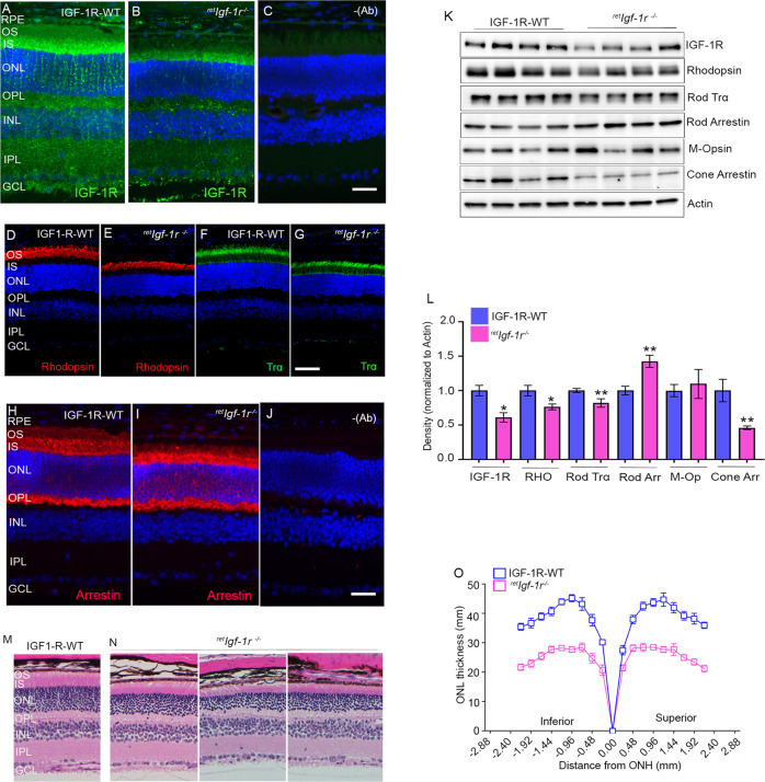
Fig. 4. Expression of IGF-1R and structural characterization of retIgf-1r−/−mice.
Prefer-fixed sections of six-week-old IGF-1R-WT and retIgf-1r−/− mouse retinas were subjected to immunofluorescence with IGF-1R (A, B), rhodopsin (D, E), rod-Trα (F, G) and rod arrestin (H, I) antibodies. (C and J) represent the omission of primary antibodies. Scale bar = 50 µm. RPE, retinal pigment epithelium; OS, outer segments; IS, inner segments; ONL, outer nuclear layer; OPL, outer plexiform layer; INL, inner nuclear layer; IPL, inner plexiform layer; GCL, ganglion cell layer. Retina lysates from the IGF-1R-WT and retIgf-1r−/− mice were subjected to immunoblot analysis with IGF-1R, rhodopsin, rod-Trα, rod arrestin, M-opsin, cone-arrestin, and actin antibodies (K). Densitometric analysis of retinal proteins normalized to actin (L). Data are mean ± SEM (n = 4). An unpaired nonparametric Mann-Whitney test was used to determine the significance. *p < 0.05; **p < 0.02. Sections from 6-week-old IGF-1R-WT (M) and retIgf-1r−/− (N) mice were hematoxylin and eosin-stained and examined for morphology. Plots of total retinal thickness were measured from the optic nerve head (ONH) in the inferior and superior regions of the retinas of 2-month-old IGF-1R-WT and retIgf-1r−/− mice (O). Data are mean ± SEM (n = 8). Two-way ANOVA was used, corrected for multiple comparisons by controlling the False Discovery Rate using a two-stage linear step-up procedure of the Benjamini, Krieger, and Yekutieli test. There was a significantly greater loss of rod nuclei in both hemispheres of the retIgf-1r−/− mouse retinas than in the IGF-1R-WT retinas (p < 0.001).

