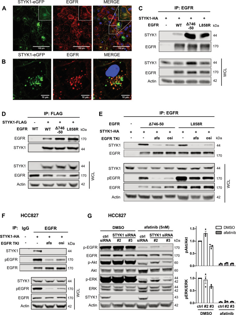
Fig. 3. STYK1 colocalizes and preferentially interacts with mutant EGFR in a manner dependent on EGFR activation.
A, B HCC827 cells were stably transduced with STYK1-eGFP. After fixation, the cells were stained using an anti-EGFR antibody. STYK1-eGFP, EGFR and nuclei (Hoechst) were visualized by Confocal microscopy. Insets show magnifications. C, D HEK293T cells were transfected with STYK1-HA or STYK1-FLAG and EGFR (wild-type (WT), Δ746-750, or L858R) and incubated for 24 hours. Cell lysates were subjected to immunoprecipitation with EGFR- (C) and FLAG- (D) antibodies. Immunoprecipitates and whole-cell lysates (WCL) were subjected to Western blotting using the indicated antibodies. E HEK293T were transfected with STYK1-HA and EGFR (wild-type (WT), Δ746-50, or L858R) and treated overnight with afatinib (10 nM) or osimertinib (10 nM). Twenty-four hours post-transfection, cell lysates were subjected to immunoprecipitation using anti-EGFR antibodies. Immunoprecipitates and WCL were subjected to Western blotting using the indicated antibodies. F STYK1-HA was transfected in HCC827 cells. After an overnight treatment with afatinib (10 nM) or osimertinib (10 nM) and a total incubation of 24 h post-transfection, lysates were prepared and subjected to immunoprecipitation using anti-EGFR or IgG control antibodies. Precipitates and WCL were immunoblotted with the indicated antibodies. G HCC827 cells were reverse transfected with control or STYK1 siRNAs, and 48 hours post-transfection 5 nM afatinib was added for an additional 24 h. Total lysates were immunoblotted with the indicated antibodies. pAkt/Akt and pERK/ERK levels were quantified with the help of the LI-COR Odyssey software. Normalized values of three independent experiments are shown as mean ± SEM.

