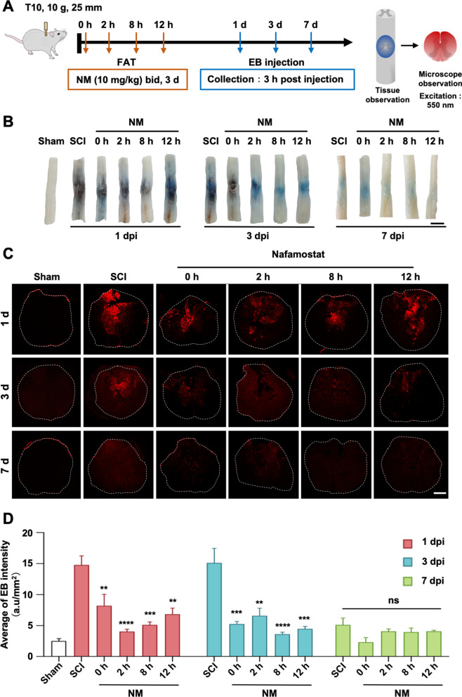
Fig. 6.
Nafamostat prevented blood–spinal cord barrier breakdown after SCI in rats. A Illustration describing the experiment design for detection of the permeability of BSCB by Evans Blue (EB) leakage. After different FAT treatment of NM (10 mg/kg, bid) for 3 days after SCI, 1 mL EB (10 mg/mL) was injected by tail vein. EB solution was allowed to circulate for 3 h before rats were sacrificed. EB leakage was detected using fluorescence microscopy (FM) under the excitation of 550-nm wavelength light. bid: bis in die. B Representative photos of general histogram of EB leakage after SCI at 1 d, 3 d, 7 d post-injury of first administration, respectively. Scale bar = 1 cm. C Representative fluorescence microscopy photographs of EB (red). Rows represent different groups, and column represents days post-injury. The border of the spinal cord is framed by white dotted line. Scale bar = 500 μm. D Quantitative analysis of average fluorescence intensity of EB in spinal cord (data shown as mean ± SEM, one-way ANOVA with Tukey's post hoc test, **P < 0.01, ***P < 0.001 vs. the SCI group, n = 4)

