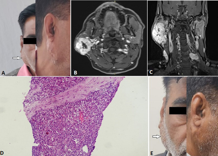
Figure 5.
A) right parotid swelling (pre-treatment) (arrow); B) MRI inT1 showing parotid involvement; C) MRI in T2 showing the tumor involvement (coronal view); D) low power magnification (x100): fibro collagenous tissue showing infiltration by a solid tumor arranged in sheets and nests; E) right parotid swelling (post-treatment) (arrow)

