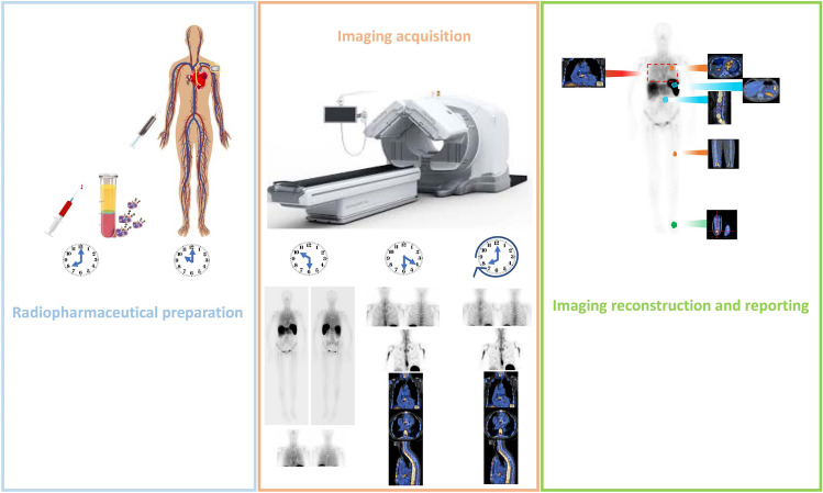
Fig. 2.
Schematic representation of the flow in radiolabeled WBC scintigraphy. First in the left panel (azul), the radiopharmaceutical preparation starting with blood sampling and the WBC isolation. After the administration of the radiolabeled cells, the patient is scanned at different time points (middle panel, orange). Early images (30 min) consists of total-body and spot of the thorax. Late images (4–6 h) and delayed images (20 h) includes spot images and SPECT/CT of the thorax, eventually followed by additional SPECT/CT based on the specific clinical condition. Finally, the images are reconstructed, reoriented, and assessed for the presence of uptake at valve/devices and extracardiac disease involvement as in case of septic embolisms, metastatic sites of infection, and the portal of entry or alternative source of infections (right panel, green)

