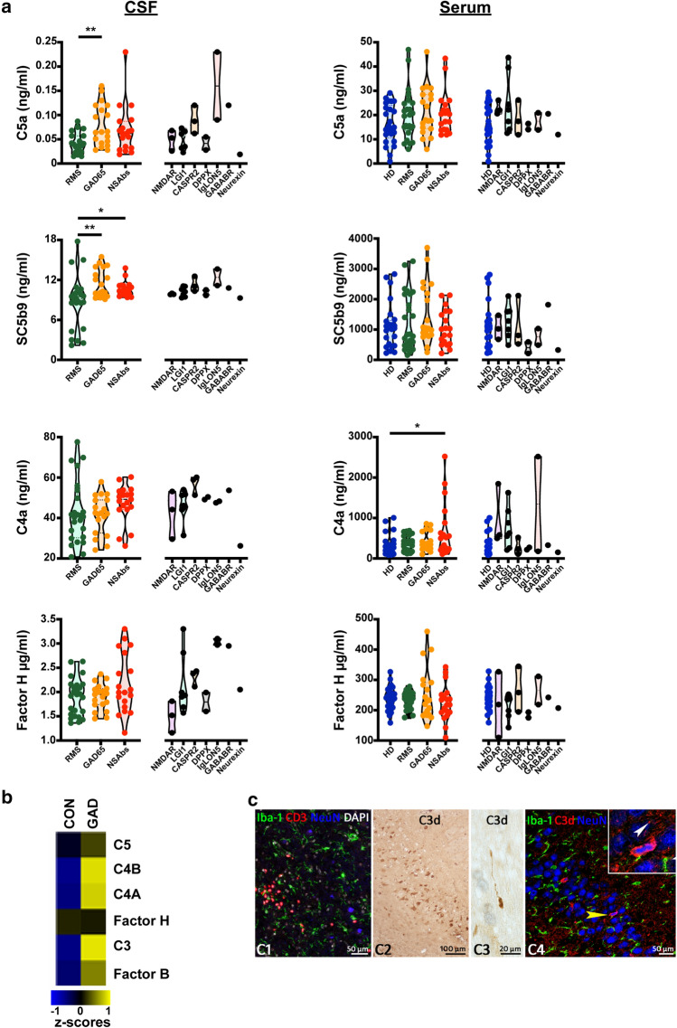
Fig. 1.
a Activated complement proteins in CSF and sera of NSAbs+ (n = 19) and GAD65-Ab+ (n = 19) encephalitis patients compared to RMS patients (n = 25) and HD (n = 25). Right panels depict levels in individual clinical entities of NSAbs-AE. *p ≤ 0.05, **p ≤ 0.001. b Microarray analysis of control (n = 7, CON) and GAD-Ab+ limbic encephalitis (n = 5, GAD) hippocampal tissue shows that mRNA expression for complement factors C3, C4A and C4B are strongly upregulated in GAD-Ab+ encephalitis. Shown are means of z-scores. (c1) Staining for T cells (CD3), Microglia (Iba-1) and neurons (NeuN) shows infiltration of T cells in the hippocampal parenchyma of a GAD-AE patient. DAPI is used as a nuclear counterstain. (c2) Immunohistochemistry for C3d shows hippocampal neurons. (c3) C3d reactivity also is seen in axonal spheroids in white matter tracts. (c4) Confocal fluorescence staining here shows NeuN+ neurons of the DG. The yellow arrowhead points toward a neuron with a condensed nucleus as enlarged in the inset. C3d staining is a representative image of one of the five patients that showed C3d reactivity

