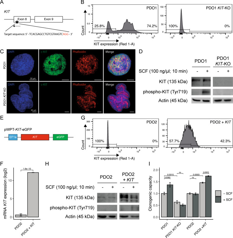
Fig. 2. Genomic engineering of KIT in patient-derived tumor organoids increases regenerative capacity.
A Schematic overview of CRISPR-Cas9 mediated KIT gene knockout in PDO1. Designed single-guide RNA targets KIT exon 8. B Flow-cytometry analyses showing deletion of cell-surface KIT expression in PDO1KIT-KO. C Immunofluorescence imaging demonstrating DAPI, c-KIT, and Phalloidin staining in PDO1CONTROL and PDO1KIT-KO, scale bar is 50 µm. D Western blot analysis of KIT and phosphorylated KIT upon stimulation with stem-cell factor (SCF, 100 ng/µl) for 10 min in PDO1CONTROL and PDO1KIT-KO. E Schematic overview of lentiviral vector to overexpress KIT, which is driven by the human EF1a promotor in PDO2. F mRNA levels of KIT expression in PDO2CONTROL and generated PDO2KIT variant. G Flow-cytometry analyses showing presence of cell-surface KIT expression in PDO2KIT. H Western blot analysis of KIT and phosphorylated KIT upon stimulation with stem-cell factor (SCF, 100 ng/µl) for 10 min in PDO2CONTROL and PDO2KIT. I Regenerative capacity of PDOs with KIT variants was assessed by counting the number of regenerated organoids. At least two independent experiments with three technical replicates. An unpaired t-test was applied to assess the significance between the groups.

