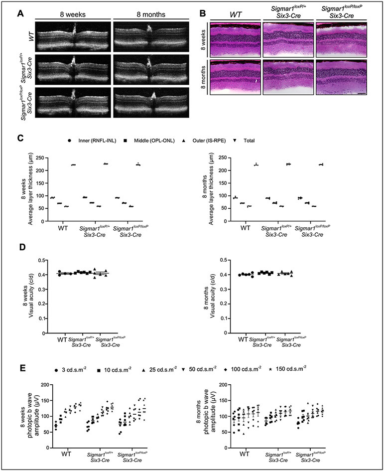
Figure 3.
Normal visual function of Sigmar1 conditional knockout mice at the early adult stage. (A) Representative optical coherence tomography (OCT) B-scan image, (B) Average thickness of retinal layers obtained from OCT images and quantified using DIVERS software including total retinal thickness (total), inner retina (RNFL, inner plexiform layer, INL); middle retina (OPL & ONL), outer retina (IS, outer segments, RPE). (C) Visual acuity measured by OMR and expressed as cycles/degree (c/d) and (D) Photopic b-wave amplitudes measured by ERG as responses to increasing light intensities expressed as candela-seconds per meter squared (cd.s.m2) were compared in wild type, Sigmar1loxP/+; Six3-Cre, and Sigmar1loxP/loxP; Six3-Cre mice at P56. RNFL: retinal nerve fiber layer; INL: inner nuclear layer; OPL: outer plexiform layer; ONL: outer nuclear layer; IS: inner segment; RPE: retinal pigment epithelium.

