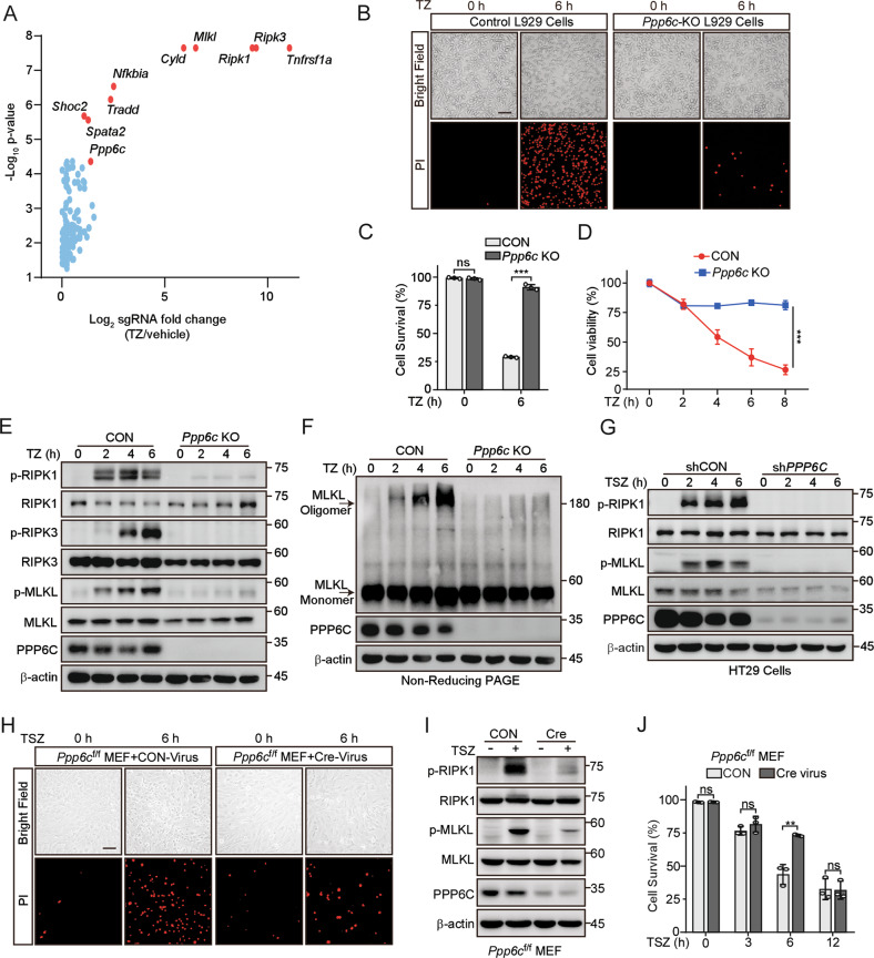
Fig. 1. Deletion of Ppp6c prevents TNF-induced necroptosis.
A Scatter diagram revealed that sgRNAs targeting Ppp6c were selected during TNF + Z-VAD-FMK (TZ) treatment in L929 cells. B, C Control and Ppp6c-KO L929 cells were treated with TZ for 6 h and analyzed by propidium iodide (PI) staining under a microscope (B) or quantified by flow cytometer (C). Scale bar, 50 μm. D Control and Ppp6c-KO L929 cells were treated with TZ for the indicated time and the cell viability was measured by CCK8. E Control and Ppp6c-KO L929 cells were treated with TZ for the indicated time and the activation of RIPK1, RIPK3 and MLKL proteins was monitored by immunoblot. F Control and Ppp6c-KO L929 cells were treated with TZ for the indicated time and cell lysates were resolved on non-reducing PAGE for immunoblot. G Control and PPP6C-knockdown HT29 cells were treated with TSZ for the indicated time and cell lysates were probed with indicated antibodies. H–J Immortalized Ppp6cf/f MEF cells were infected with control lentivirus or Cre-Lentivirus for 72 h and followed by the treatment with TSZ for 6 h. Cells were stained with PI and analyzed under a microscope (H) or lysed for immunoblot with indicated antibodies (I), or quantified by flow cytometer after PI staining (J). Scale bar, 50 μm. Data shown are representative of three independent experiments and presented as means ± SDs of triplicates (C, D, J). **p < 0.01, ***p < 0.001, with an unpaired Student’s t-test (C, D, J).

