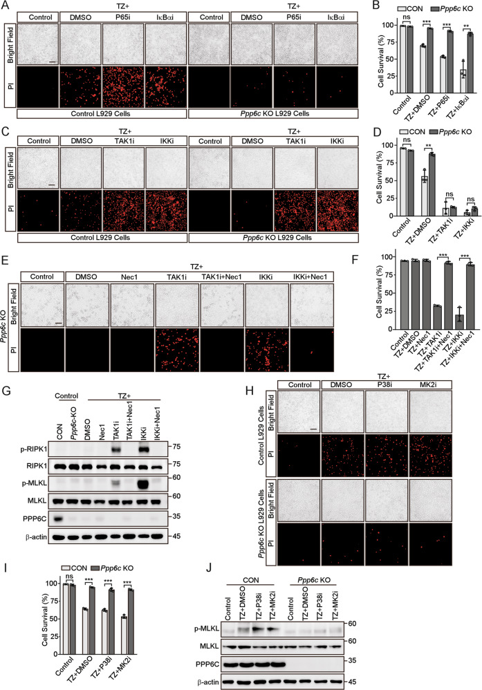
Fig. 3. Inhibition of TAK1 or IKK activity restores TNF-induced necroptosis in Ppp6c deficient L929 cells.
A, B Control and Ppp6c-KO L929 cells were pretreated with P65 inhibitor (Maslinic acid, 20 μM) or IκBα inhibitor (BAY117085, 10 μM) for 30 min and then treated with TZ for 4 h. Cells were stained with PI, and analyzed under a microscope (A) or quantified by flow cytometer (B). Scale bar, 50 μm. (C, D) Control and Ppp6c-KO L929 cells were pretreated with TAK1 inhibitor (5Z-7-Oxozeaenol, 1 μM) or IKKα/β inhibitor (IKK16, 1 μM) for 30 min and then treated with TZ for 4 h. Cells were stained with PI, and analyzed under a microscope (C) or quantified by flow cytometer (D). Scale bar, 50 μm. E–G Ppp6c-KO L929 cells were pretreated with TAK1 inhibitor (5Z-7-Oxozeaenol, 1 μM) or IKKα/β inhibitor (IKK16, 1 μM) in the presence or absence of Necrostatin-1 (10 μM) for 30 min, and then treated with TZ for 4 h. Cells were stained with PI and analyzed under a microscope (E) or quantified by flow cytometer (F). Cell lysates were probed with indicated antibodies (G). Scale bar, 50 μm. H–J Control and Ppp6c-KO L929 cells were pretreated with P38 inhibitor (Adezmapimod, 10 μM) or MK2 inhibitor (MK2-IN-1, 10 μM) for 30 min, and then treated with TZ for 4 h. And cells were stained with PI and analyzed under a microscope (H) or quantified by flow cytometer (I), and cell lysates were probed with indicated antibodies (J). Scale bar, 50 μm. Data shown are representative of three independent experiments and presented as means ± SDs of triplicates (B, D, F, I). **p < 0.01, ***p < 0.001, with an unpaired Student’s t-test (B, D, I) or one-way ANOVA analysis (F).

