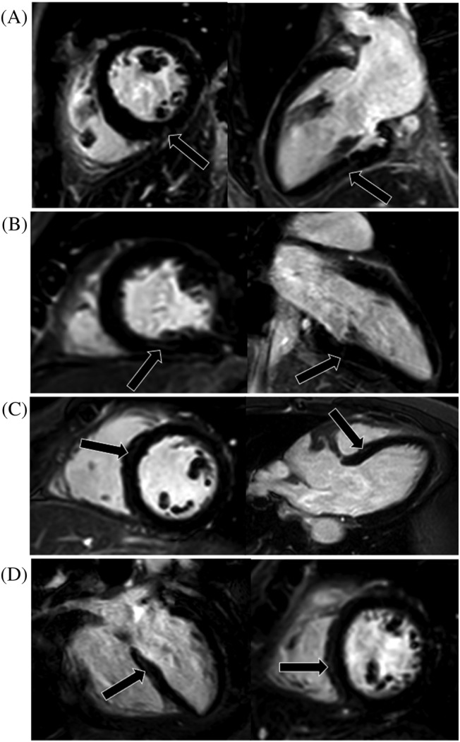
Figure 2.

The illustration of all four LGE positive patients' myocardial injury. (A–D) represents cases 1–4, respectively. One short axis and orthogonal long‐axis PSIR images showed focal LGE positive (black arrows) for each patient at the basal‐mid level of the left ventricular in inferior or inferoseptal segments. LGE was most commonly seen in the subepicardial location (A, C, and D). LGE, late gadolinium enhancement; PSIR, phase sensitive inversion recovery.
