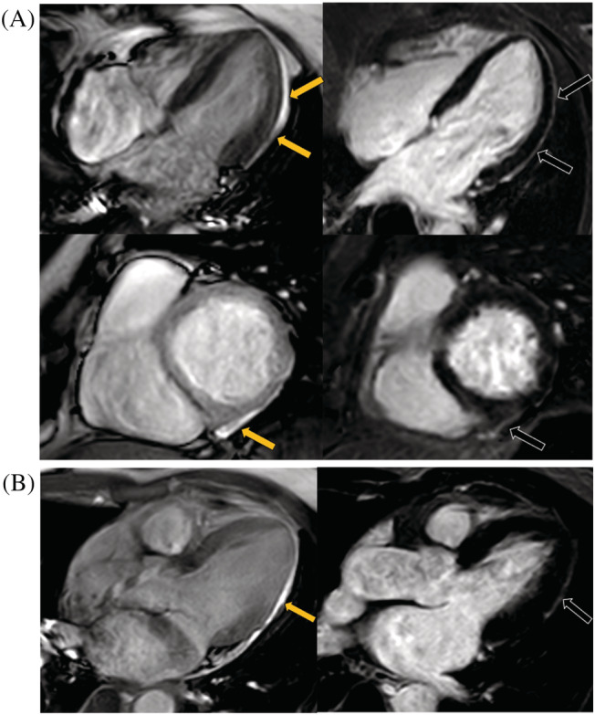
Figure 3.

The illustration of all two LGE positive patients' pericardium injury. (A, B) represents cases 1–2, respectively. Cine images showed pericardial effusion at LV free wall (yellow arrows), and PSIR images showed the corresponded pericardial enhancement (black arrows) for each patient. LV, left ventricular; PSIR, phase sensitive inversion recovery.
