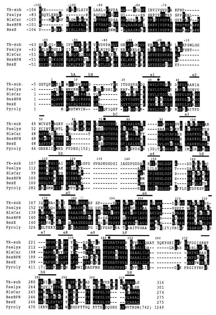
FIG. 1.
Alignment of subtilisin sequences. The amino acid sequence of T. kodakaraensis subtilisin (Tk-sub) is compared with those of P. aerophilum aerolysin (Paelys) (accession no. S76079), B. licheniformis subtilisin Carlsberg (BlsCar) (accession no. X03341), B. amyloliquefaciens subtilisin BPN′ (BasBPN) (accession no. X00165), B. subtilis subtilisin E (BssE) (accession no. K01988), and pyrolysin core (Pyroly) (accession no. U55835). Gaps are denoted by dashes. The numbers in parentheses represent the numbers of the amino acid residues inserted or extended at the positions indicated. The conserved amino acid residues are denoted with white letters. The amino acid residues that form a catalytic triad and the asparagine residue that forms an oxyanion hole are denoted by solid and open circles, respectively. The numbers represent the positions of the amino acid residues starting from the N terminus of the mature proteins for bacterial subtilisins and pyrolysin and the positions of putative catalytic domains for T. kodakaraensis subtilisin and aerolysin. The eight α-helices (hA to hH) and nine β-strands (e1 to e9) of subtilisin BPN′ are shown above the sequences.

