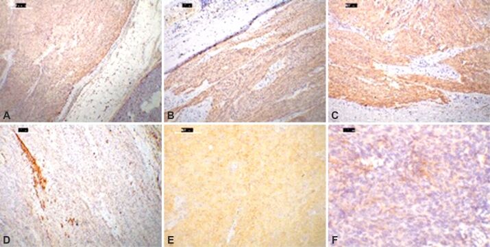
Figure 3.
Immunohistochemical stainings of diverse EGIST locations: (A) Micrograph with anti-CD34 antibody moderate immunostaining in tumor cells and vascular channels in the nearby adipose tissue and pancreas, while anti-CD117 antibody (B) and anti-DOG1 antibody (C) show diffuse, strong positivity and scattered tumor cells and fascicles for α-SMA (D); (E) Micrograph of a mesentery-located epithelioid EGIST, DOG1 negative (not showed), PDGFRA positive; (F) EMA can be focally and patchy expressed in a retroperitoneal spindle cell-shaped EGIST. DAB, Hematoxylin counterstaining: (A–C) ×40; (D) ×100; (E) ×200; (F) ×400. α-SMA: Alpha-smooth muscle actin; CD: Cluster of differentiation; DAB: 3,3’-Diaminobenzidine; DOG1: Discovered on gastrointestinal stromal tumor (GIST) 1; EGIST: Extragastrointestinal stromal tumors; EMA: Epithelial membrane antigen; PDGFRA: Platelet-derived growth factor receptor alpha

