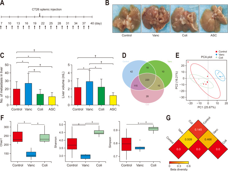
Fig. 1.
Changes in gut microbiota and colorectal cancer (CRC) liver metastasis induced by different antibiotics. (A) Schematic diagram of the mouse experimental process of CRC liver metastasis with different antibiotics (upper arrow: CRC liver metastasis model, as established by colon 26 [CT26] splenic injection; lower arrow: antibiotic treatment). (B) Image of CRC liver metastases in animals treated with different antibiotics at the end of the experiment. (C) Number of CRC liver metastases and liver volume in different groups (n=15 per group; a mouse in the Vanc group and a mouse in the ASC group were lost during the experiment due to emaciation and intestinal obstruction). (D) Venn diagram of the total number of species among the control, Vanc, and Coli groups (n=6 per group). (E) Principal coordinates analysis (PCA) of operational taxonomic units in the control, Vanc, and Coli groups. (F) Alpha diversity analysis of the gut microbiomes among the control, Vanc, and Coli groups. Alpha diversity includes the Shannon, Chao1, and Simpson indices. (G) Beta diversity, reflected by the weighted Unifrac distance.
Control group, untreated; Vanc group, vancomycin; Coli group, colistin; ASC group, a mix of ampicillin, colistin and streptomycin. *p<0.05, †p<0.01.

