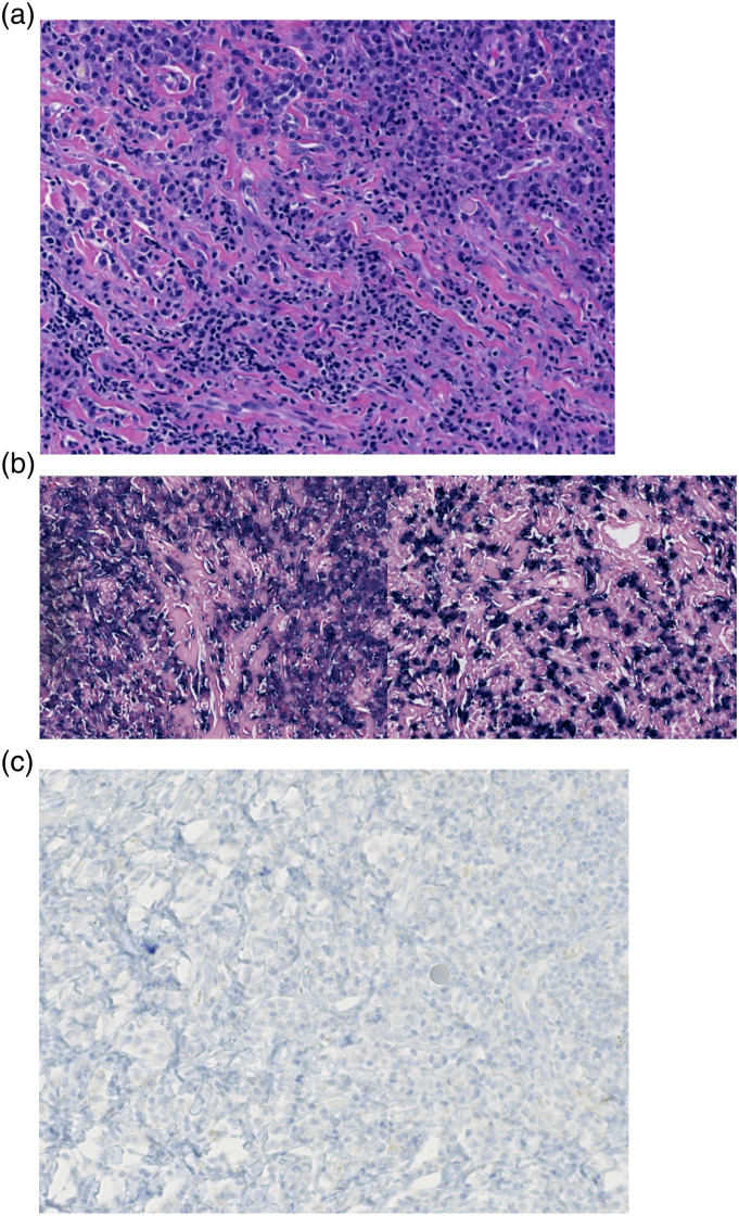
Figure 1.
(a) H&E stain reveals a well-circumscribed mass composed of numerous plasma cells and lymphocytes in a background of dense fibrosis. (b) In-situ hybridization for kappa (left) and lambda (right) light chains shows a polyclonal mix of plasma cells. (c) An immunostain for ALK-1 is negative and ALK FISH is negative for a translocation.

