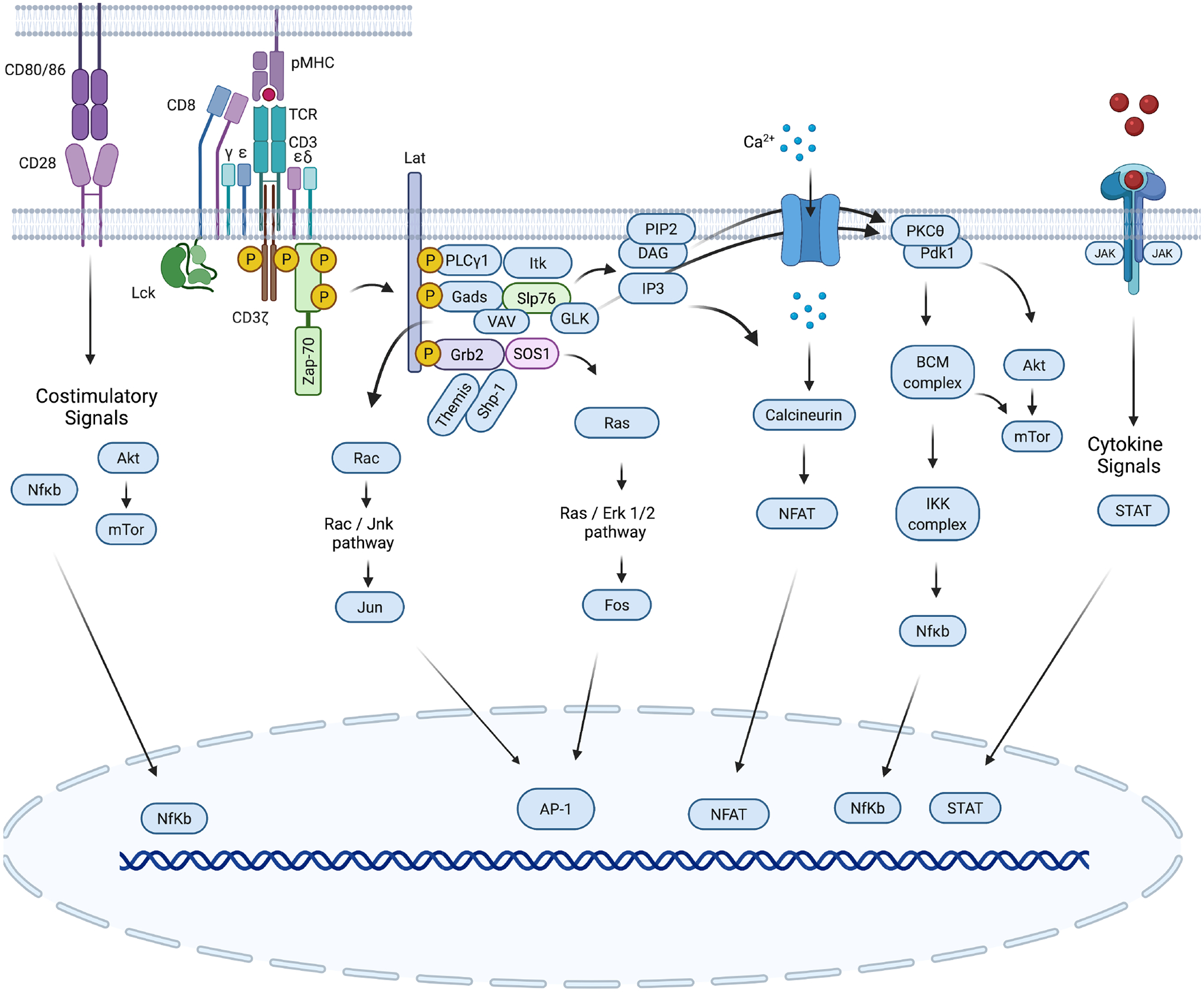
Fig. 2. Canonical TCR signaling.

The TCR is composed of an αβ heterodimer that noncovalently associates with the CD3εδ and CD3εγ heterodimers and a ζ-chain homodimer (ζζ). Binding of the TCR to peptide-MHC complexes on the antigen-presenting cell (APC) stimulates TCR signaling. The co-receptor CD8 or CD4 interacts with the pMHC and approaches the TCR, enabling the kinase Lck to phosphorylate tyrosines within the ITAM motifs of the CD3 chain tails (10 in total). The kinase ZAP70 is then recruited to the TCR/CD3 complex through its association with phosphorylated ITAMs, and its phosphorylation by Lck causes its activation. Zap-70, in turn, phosphorylates the adapter proteins Slp76 and Lat, which form a signalosome that is a hub for other kinases and substrates to distribute and diversify the TCR signal. The enzyme PLCγ1 is activated by the kinase Itk and mediates the generation of diacylglycerol (DAG) and inositol 1,4,5-trisphosphate (IP3). Both molecules act as second messengers for two important signaling cascades. IP3 initiates calcium (Ca2+) signaling, which leads to the activation of the transcription factor NFAT, whereas DAG and Ras-GRP enables activation of the Ras-ERK1/2 cascade (together with Grb-2–Sos). DAG, Slp76/Vav, and PI3K activity cooperate in the activation of the kinase PKCθ, which induces the formation of the CBM complex (Carma1/Bcl10/MALT1), which is followed by activation of the IKK complex (IKKα-IKKβ-IKKγ), leading to the nuclear translocation of the transcription factor NF-κB. The activity of the kinase PI3K, which is activated by TCR signals, can be amplified by costimulatory CD28 signals and enables the localization of the kinases PDK1 and Akt at the plasma membrane, where Pdk1 activates Akt. Subsequently, Akt regulates mTOR signaling, which modulates T cell metabolism. TCR engagement leads to an increase in the concentration of intracellular Ca2+, activation of the CBM complex and mitogen-activated protein kinase pathways (mediated by ERK and JNK), which ultimately results in the activation and nuclear translocation of the transcription factors NFAT, NF-κB, and AP-1.
