Fig. 2.
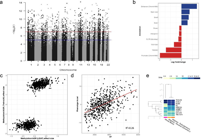
Subcutaneous adipose tissue differential methylation signature of VF. a A Manhattan Plot of the associations between VF and DNA methylation, taking into account BMI resulting from epigenome-wide association analysis (N = 538). The red line shows the multiple testing threshold (P = 1.14 × 10⁻⁷), and the blue line shows a relaxed significant threshold (P = 1 × 10⁻⁵). b Enrichment and depletion of VF-DMPs resulting from epigenome-wide association analysis (N = 538) across genomic annotations. Log Fold changes show the proportion of VF-DMPs annotated to particular genomic annotations, compared to all CpGs tested in each annotation. The plots show only annotation categories with significant enrichments and depletions. c Comparison between the discovery cohort (TwinsUK) (N = 538) and validation cohort (LEAP; N = 104) showing effect size for the associations resulting from the regression analysis between AGR and methylation at VF-DMPs without adjustment for cell composition in LEAP. d Significant positive association between PhenoAge acceleration and VF accumulation with a line of best fit shown in red along with its R2 value (N = 538). e GTEx gene expression levels in whole blood, visceral fat and subcutaneous fat for the 9 genes identified in the study following the omic integration showing shared expression levels in the two types of adipose tissue (SAT and VAT), but differences in expression levels between adipose tissue and whole blood. TPM is transcripts per kilobase million, and the median expression levels are shown in Additional file 2: Table S10

