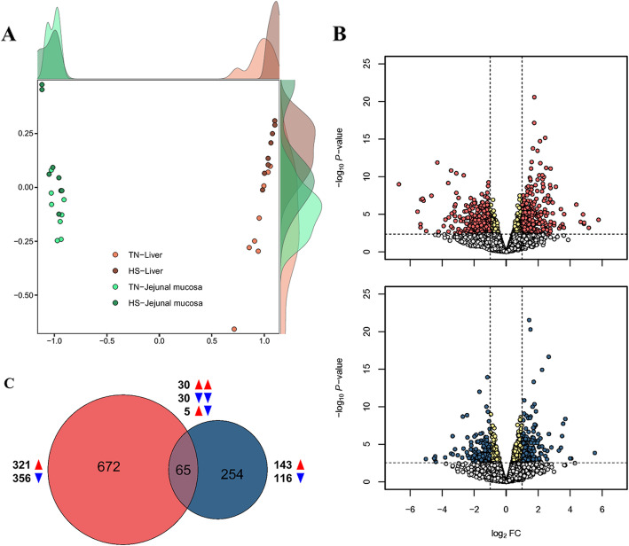
Fig. 2.
Overall transcriptomes in liver and jejunal mucosa. A Multidimensional scaling (MDS) reveals separate clusters between the two tissues based on the transcriptomes under thermoneutral zone (TN) and heat stress (HS). B Volcano plots indicating significant differently expressed genes (DEGs) in the liver (red) and jejunal mucosa (blue). The x and y axes of the volcano plots show the log2 fold changes and -log10 P-values, respectively. C Venn diagram is visualized based on overlapping number of DEGs between liver (red) and jejunal mucosa (blue) tissues

