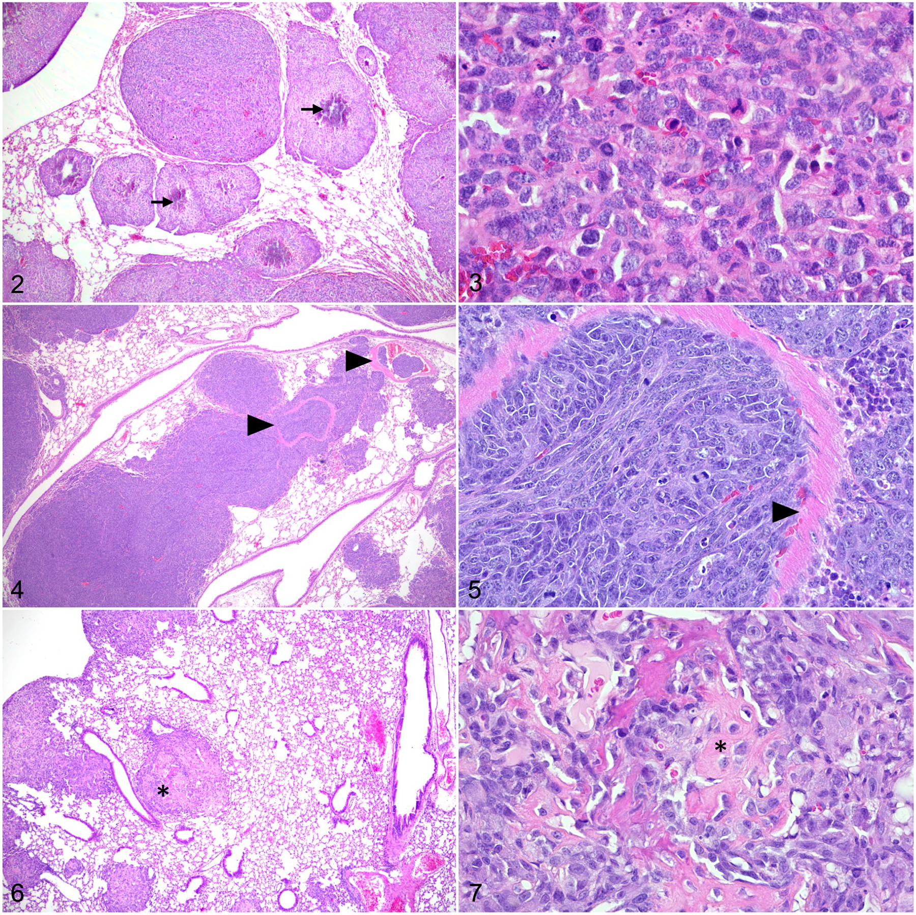
Figures 2–7.

Osteosarcoma, lung, mouse. Hematoxylin and eosin. Figure 2. Following para-tibial injection, SaOS-2 cells form pulmonary nodules with central mineralization (arrows). Figure 3. Lung metastasis of SaOS-2 cells with mitotic figures. Figure 4. Following tail vein injection, DLM8 murine OS cells form numerous pulmonary nodules. Tumor emboli are observed within pulmonary vessels (arrowheads). Figure 5. The remnants of a vessel wall (arrowhead) within a DLM8 cell tumor nodule. Figure 6. Para-tibial injection of MC-KOS canine OS cells forms multiple pulmonary tumors. Few tumor nodules contain central deposits of eosinophilic matrix (asterisk). Figure 7. Lung metastasis of MC-KOS cells with production of variably mineralized osteoid (asterisk).
