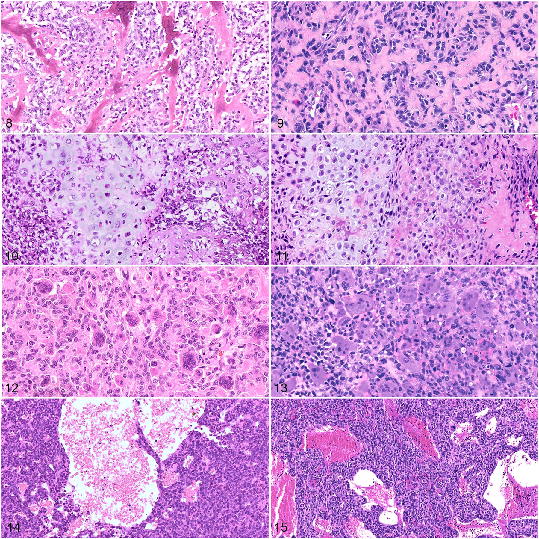
Figures 8–15.

Osteosarcoma, dog (left column) and human (right column). Hematoxylin and eosin. Figures 8 and 9. Osteoid is a distinguishing tumor feature observed in canine (Figure 8) and human (Figure 9) osteosarcomas. Figures 10 and 11. Some canine (Figure 10) and human (Figure 11) osteosarcomas also contain chondroid matrix (chondroblastic subtype). Figures 12 and 13. Giant cell-rich osteosarcomas are an uncommon histologic variant reported in both dogs (Figure 12) and humans (Figure 13). Figures 14 and 15. Telangiectatic osteosarcomas are histologically characterized by blood-filled spaces lined by tumor cells (dog, Figure 14; human, Figure 15).
