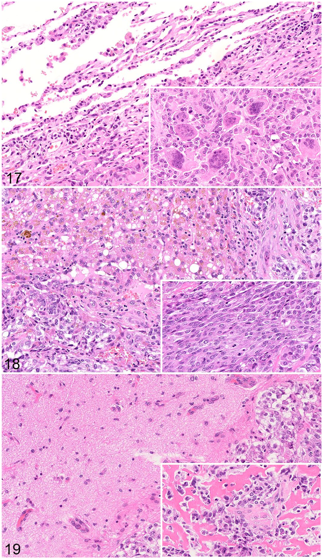
Figures 17–19.

Metastatic osteosarcoma, dog. Hematoxylin and eosin. Figure 17. Lung. Alveoli along the edge of the metastasis are compressed; many contain erythrocytes. Inset: multinucleated cells are observed throughout the mass. Figure 18. Liver. There is mild lymphocytic infiltration of the hepatic parenchyma adjacent to the tumor. Many hepatocytes are vacuolated and contain brown pigment. Inset: The metastasis is comprised of polygonal to spindle-shaped cells. Figure 19. Brain. There is a well-demarcated OS metastasis with compression of the adjacent parenchyma. Inset: Osteoid is abundant within the tumor.
