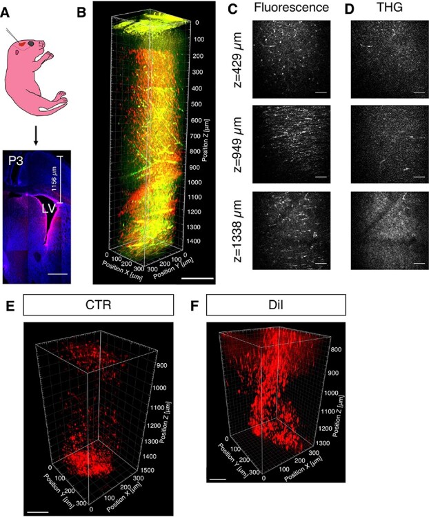
Figure 1.

Validation of 3PM in postnatal SVZ imaging. (A) A schematic of LV injection in postnatal day 1 (P1) CD1 pup and tiled confocal imaging of a representative brain section labeled with CTO (red) and DAPI (blue), 2 days post injection (P3). (B) 3D reconstruction from 0 to 1472 μm below the pial surface of pup with CTO (3 dpi; red, CTO fluorescence; green, THG). (C, D) Selected XY frames at different depths in (B) (see Supplementary Movie 1). (E) 3D reconstruction of white matter and SVZ of pups injected with CTR (3 dpi). (F) 3D reconstruction of white matter and SVZ of pups injected with DiI (5 dpi). Scale bars represent 500 μm in A, 200 μm in B, 50 μm in C and D, 100 μm in E; 80 μm in F.
