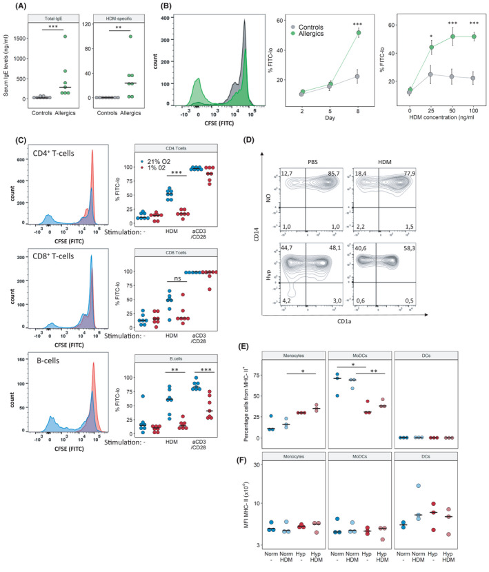
FIGURE 5.

The human adaptive immune response to HDM is impaired under hypoxic conditions. Immune cells were isolated from peripheral blood of HDM‐allergic patients or healthy donors. PBMCs were stimulated in vitro with 25, 50 or 100 µg HDM or aCD3/28 beads under normoxia (Norm) and hypoxia (Hyp) for 8 days. (A) Total IgE and HDM‐specific IgE measured by ELISA from serum of blood donors. (B) CD4+ T‐cell proliferation in response to HDM stimulation was measured by flow cytometry (non‐allergic vs. allergic subjects, Norm only). Representative CFSE histogram (left), proliferation response over time (middle), proliferation with increasing HDM concentrations at day 8 post‐stimulation (right). (C) Flow cytometry analysis of cell proliferation on day 8 as % CFSE‐lo of T and B cells, either unstimulated (‐), in response to 100 µg/ml HDM or aCD3/CD28 stimulation. (D) Flow cytometry analysis of antigen‐presenting cells (APCs) on day 8 as % of CD45+MHCII+. Monocytes (CD14+CD11b+CD1a−), MoDCs (monocyte‐derived dendritic cells, CD14+CD11b+CD1a+), DCs (dendritic cells, CD1a+CD11b+CD14−). (E) Quantification of subpopulations of APCs stimulated with or without HDM under Norm or Hyp. (F) Mean fluorescent intensity (MFI) of MHCII in populations from (E). Full gating strategy is given Figures [Link], [Link]. n = 7 (A–C), n = 3 (D–F) p < .05, **p < .01
