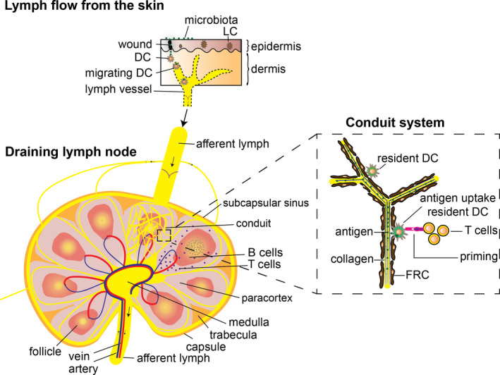
FIGURE 1.

Schematic view of T‐cell activation in a draining lymph node after cutaneous infection. Migrating DC take up microbial antigens at the wound site and route via a lymphatic vessel to a local draining lymph node. A draining lymph node consists of micro‐domains containing paracortical T‐cell areas and follicular B‐cell areas. Lymph fluid (less than 70 kDa) enters the conduit system, which forms a tube system within T‐cell and B‐cell areas. The conduit system consists of organized collagen fibres with FRC wrapped around it. Resident DC within T‐cell areas are able to pick up antigens that flow through the conduit system. Resident mature DC present antigen to local naïve T cells, resulting in T‐cell priming and activation
