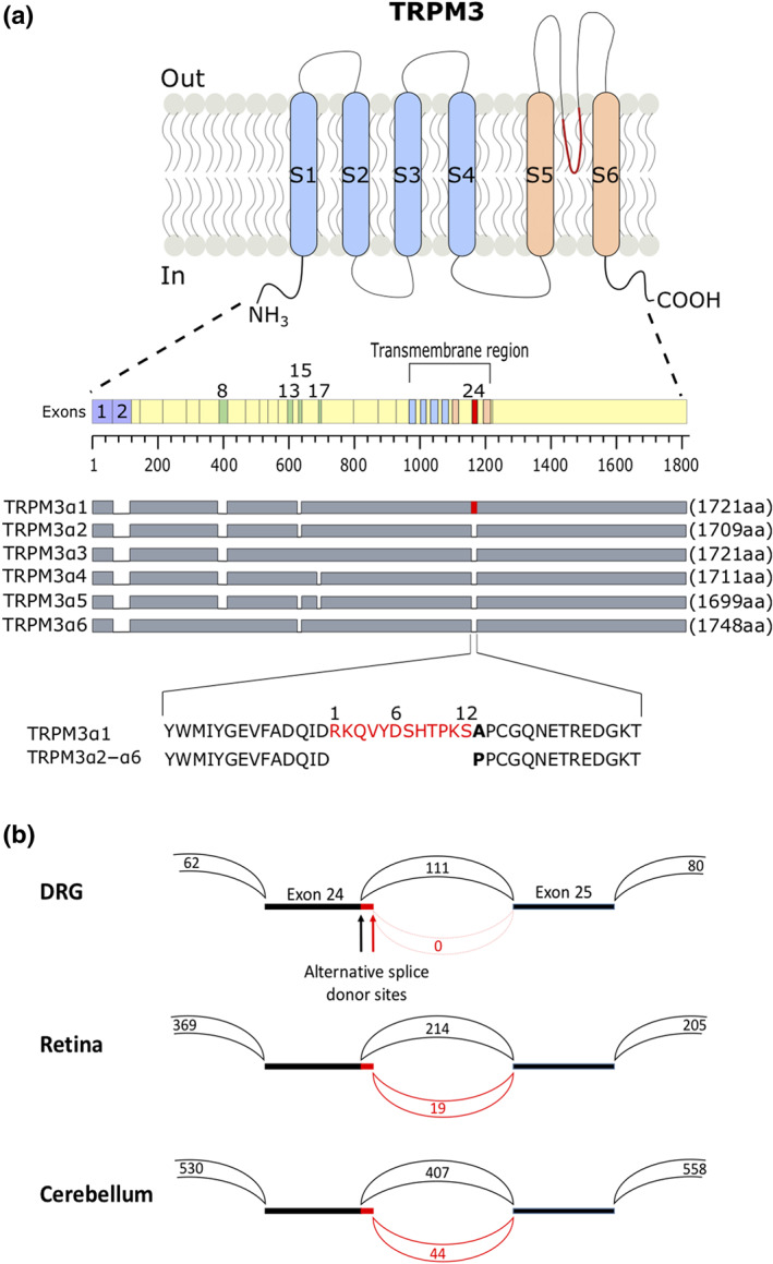
FIGURE 1.

Alternative splicing resulting in short pore and long pore TRPM3 variants. (a) Cartoon illustrating the protein differences in TRPM3α variants. Transmembrane segments S1–S4 of the voltage‐sensing domain and S5–S6 of the pore domain are illustrated in light blue and light orange; alternative exons are numbered. Protein length of different TRPM3α variants and their spliced exons are indicated as grey bars. Marked in red is the 12 amino acid stretch located in the pore loop on exon 24 that is unique to the TRPM3α variant TRPM3α1. A segment of the pore loops of TRPMα1–α6 is represented as amino acid sequence. Numbers above the sequences are referring to the 12 amino acid insertion within exon 24. (b) Sashimi plot showing the junctions between exons 23 and 25. The two alternative splice donor sites at the 3′ end of exon 24, which give rise to the short and the long pore variants, are indicated. The numbers indicate the total number of reads split across splice junctions
