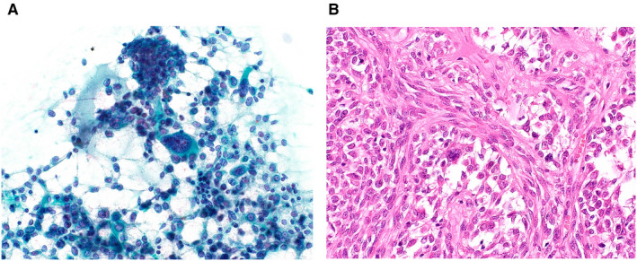
Figure 3.

(A) Cytological features of a pleomorphic adenoma that was categorized as salivary gland neoplasm of uncertain malignant potential. Many myoepithelial cells and mucin can be seen. Among these, a few large, atypical cells are present (Papanicolaou stain, original magnification ×400). (B) Histology of the resected specimen reveals cellular pleomorphic adenoma in which interspersed, bizarre myoepithelial cells can be seen (H&E stain, original magnification ×400).
