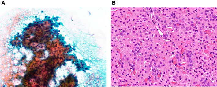
Figure 4.

(A) Cytological features of a case of nodular oncocytic hyperplasia that was categorized as salivary gland neoplasm of uncertain malignant potential. Large oncocytic cell clusters with polygonal eosinophilic cytoplasm can be seen (Papanicolaou stain, original magnification ×400). (B) The histologic specimen shows hyperplasia with ductal components (H&E stain, original magnification ×200).
