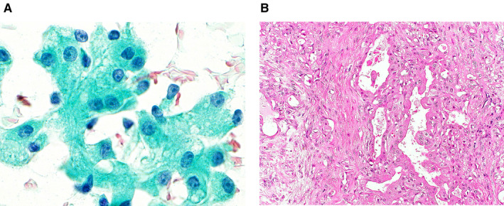
Figure 5.

(A) Cytological features of a pleomorphic adenoma that was misdiagnosed as acinic cell carcinoma. Acinar‐like clusters of epithelial cells with large amounts of lacy and granular cytoplasm and distinct nucleoli can be seen (Papanicolaou stain, original magnification ×1000). (B) On histology of the resected specimen, oncocytic changes in the ductal epithelium (right) can be seen in this typical pleomorphic adenoma (left) (H&E, original magnification ×200).
