Figure 1.
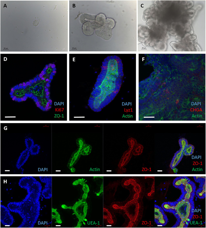
3D enteroids cultivated from bovine intestinal tissue. Representative images showing crypts isolated from bovine ileal tissue and maintained in a Matrigel dome with IntestiCult medium, from 5 independent animals. (A–C) Brightfield images of 3D enteroids cultured for 24 h (A), 3 days (B) and 7 days (C) demonstrating that they bud and proliferate over time. By 7 days the enteroid lumen fills with debris from cells sloughed off and are ready to be passaged. (D–H) Confocal images of 3D bovine enteroids stained for epithelial cell fate markers. (D) Nuclei (DAPI, blue), tight junctions (ZO-1, green) and proliferative cells (Ki-67, red). (E) Nuclei (DAPI, blue), actin (Phalloidin, green) and Paneth cells (lysozyme, red). (F) Nuclei (DAPI, blue), F-actin (Phalloidin, green) and enteroendocrine cells (chromogranin A, red). (G) Split panel of enteroids stained for nuclei (DAPI, blue), F-actin (Phalloidin, green), and tight junctions between cells (ZO-1, red). (H) Split panel of enteroids stained for nuclei (DAPI, blue), glycolipids (UEA-1, green) and tight junctions (ZO-1, red). Scale bar = 50 μm.

