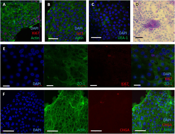
Figure 2.
2D epithelial monolayers cultured on collagen matrix. Single cells were seeded onto collagen coated wells. The 2D monolayers were cultured with IntestiCult containing the relevant inhibitors and maintained for up to 10 days. (A–F) Representative confocal microscopy images of 2D monolayers cultured on collagen coated wells from 3 separate amimals, demonstrating presence of specific cell marker proteins. (A) F-actin (phalloidin, green), nuclei (DAPI, blue) and proliferative cells (Ki67, red). (B) Nuclei (DAPI, blue), F-actin (Phalloidin, green) and Paneth cells (lysozyme, red). (C) Nuclei (DAPI, blue) and glycolipids (UEA-1, green). (D) 2D monolayer imaged by brightfield microscopy stained for Periodic Acid Schiff to show mucins produced by goblet cells. (E) Split panel of monolayer stained for nuclei (DAPI, blue), tight junctions between cells (ZO-1, green), and proliferative cells (Ki-67, red). Scale bar = 20 μm. (F) Split panel of monolayer stained for nuclei (DAPI, blue), F-actin (Phalloidin, green) and enteroendocrine cells (chromogranin A, red). Scale bar = 50 μm.

