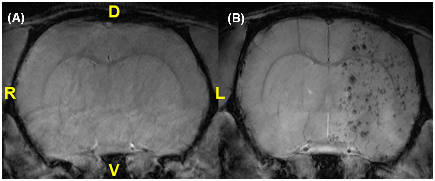
FIGURE 1.

FLASH images of in vivo rat brains following PBS (A) or 2D hMSC (B) administration. Anatomical references are provided for dorsal (D), ventral (V), left (L), and right (R) orientations, with the left anatomical hemisphere corresponding to the ipsilateral (ischemic) side and showing decoration by MPIO-labeled 2D hMSC injected intra-arterially
