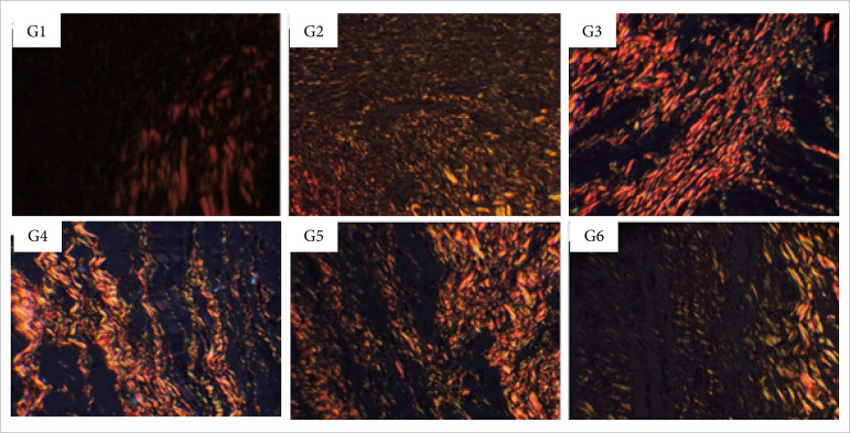
Figure 12. Collagen fibers deposition in partial thickness burn wounds experimentally induced in Wistar rats 30 days after the injury induction (DAI). Collagen fibers type I are shown in red and type III in green. Stain Picrosirius red. Augmentation: 20x scale in μm.
G1: control group with animals treated with NaCl 0.9%; G2: animals treated with 1% silver sulfadiazine; G3: animals treated with Debrigel™; G4: animals treated with Safgel™; G5: animals treated with Dersani Hydrogel™; G6: animals treated with Solosite™.

