FIGURE 1.

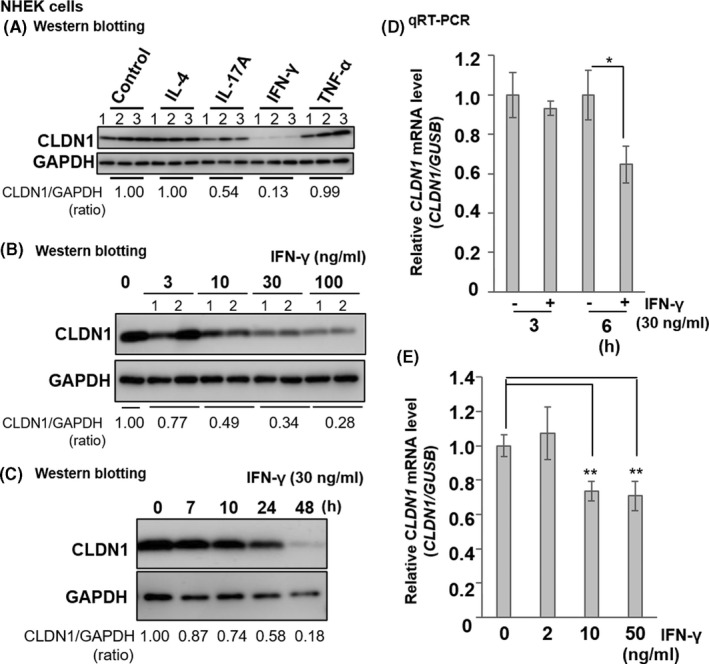
Dose‐ and time‐dependent inhibition of CLDN1 expression by IFN‐γ in normal human epidermal keratinocytes (NHEKs). (A) Effects of IL‐4, IL‐17A, IFN‐γ and TNF‐α (30 ng ml−1) on CLDN1 expression in NHEK cultures incubated with the cytokines in a 24‐well plate for 48 h. Samples were prepared from another three wells (n = 3). (B) Dose‐dependent inhibition of CLDN1 expression by IFN‐γ (3–100 ng ml−1) in NHEKs cultured with or without IFN‐γ for 48 h (n = 2). (C) Time‐dependent inhibition of CLDN1 expression in NHEKs treated with 30 ng ml−1 IFN‐γ for 0–48 h (n = 1). Western blotting performed with anti‐CLDN1 and anti‐GAPDH antibodies. Examples of typical blots in duplicate are illustrated. The expression level of CLDN1 was quantified by densitometry and normalized to GAPDH; the ratio is shown directly under each blot. Non‐treated control (0) is 1.00. The statistical analysis is shown in Figure S1. The results are representative of more than three independent experiments. (D) Changes in the relative CLDN1 mRNA expression in NHEKs cultured in a 24‐well plate with or without IFN‐γ (30 ng ml−1) for 3 or 6 h. (E) Changes in the relative CLDN1 mRNA expression in NHEKs treated with 2–50 ng ml−1 IFN‐γ for 6 h. mRNA expression was measured using qRT‐PCR. Data are presented as the mean ± SD (n = 4). Data were analysed using the Dunnett's test (*p < 0.05; **p < 0.01 vs. control (0 ng ml−1))
