
FIGURE 5.
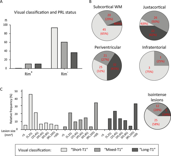
Small subcortical and periventricular lesions, and lesions that do not develop paramagnetic rims, are more likely to have T1 times associated with remyelination when evaluated a median of 4.2 years (range: 0.3–25) after lesion formation. (A) Paramagnetic rim lesions (PRL; n = 23) were nearly always categorized as “long‐T1” or “mixed‐T1” (chi‐squared test, p < 0.001). (B) Lesion location affects the relative proportions of lesion types (chi‐squared test, p = 0.02): long‐T1 lesions are most common in periventricular and juxtacortical regions, whereas short‐T1 lesions predominate in subcortical white matter but are also common in the periventricular region. (C) Histogram of sizes of lesions that were T1‐hypointense at the time of gadolinium enhancement showing that smaller lesions were more likely to evolve into “short‐T1” lesions, suggesting remyelination. Pie chart shows the fate of lesions that were T1‐isointense at the time of gadolinium enhancement; only 5 (14%) of these 42 lesions were in the long‐T1 group at follow‐up 7 T MRI. [Color figure can be viewed at www.annalsofneurology.org]