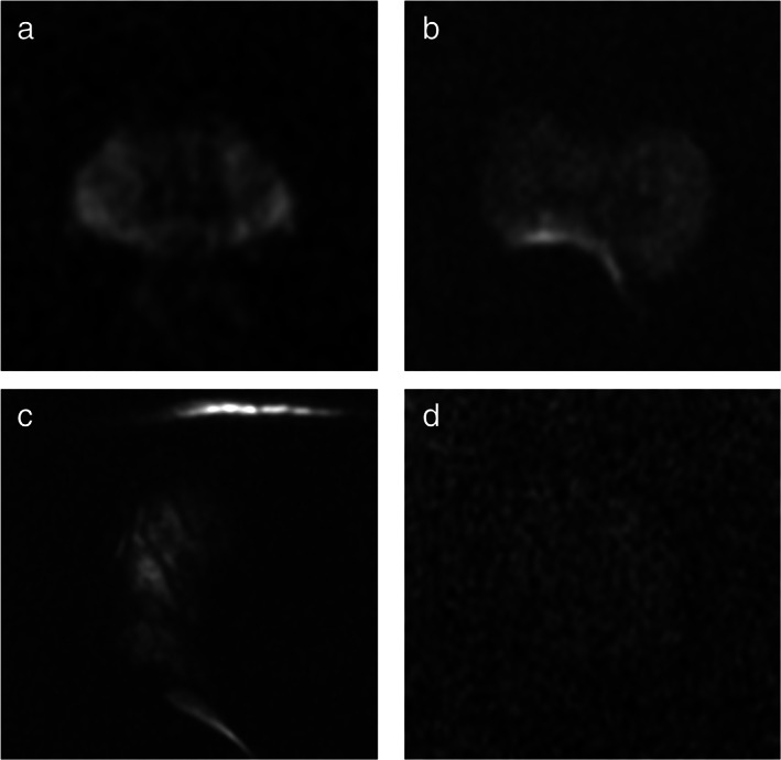
FIGURE 3.

Case examples of high‐ and low‐quality scans on DWI images. It shows examples of high‐ and low‐quality DWI images: (a) high‐quality DWI image (Q1), with good S/N ratio and no evident artifacts; (b) low‐quality image (Q0) with susceptibility artifacts caused by the presence of air in the rectum—the ability to detect foci in the right posterior peripheral zone is significantly impaired—the sequence could be repeated following attempts to expel the air from the rectum; (c) inadequate acquisition (Q0) with marked distortion and signal void due to magnetic susceptibility artifacts caused by a femoral prosthesis; (d) low‐quality image (Q0) because of inadequate S/N ratio that lower significantly the diagnostic power—the sequence needs to be repeated following optimization of the acquisition parameters.
