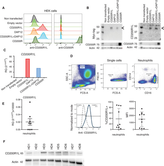
FIGURE 1.

Anti‐CD200R1L antibody specificity and CD200R1L expression in human healthy donor neutrophils. (A–C) HEK293T transfected with DNA vectors expressing indicated proteins, and analysed by (A) flow cytometry, (B) Western blot, or (C) qPCR. (A) n = 3 from 3 independent experiments, one representative experiment is shown. Gating strategy available in Supplementary Fig. S1A. (B) Samples were stained using anti‐MYC antibody (left side) or anti‐CD200R1L (right side). Arrows depict CD200R1L bands. n = 2 from 2 independent experiments. (C) n = 2 from 2 independent experiments. (D–F) Expression of CD200R1L assessed by (D) flow cytometry, (E) qPCR, and (F) Western blot. (D) A representative FACS staining indicating CD200R1L expression in neutrophils from a healthy donor. Quantification indicate the % and MFI of CD200R1L positive neutrophils. 10 different donors were analysed in 5 independent experiments (E) CD200R1L expression in purified healthy donor neutrophils by qPCR analysis, n = 7 from 3 independent experiments (F) and by Western blot n = 8 from 4 independent experiments. Actin and CD200R1L were probed on the same membrane.
Full‐length blots are included in the Supplementary Information
