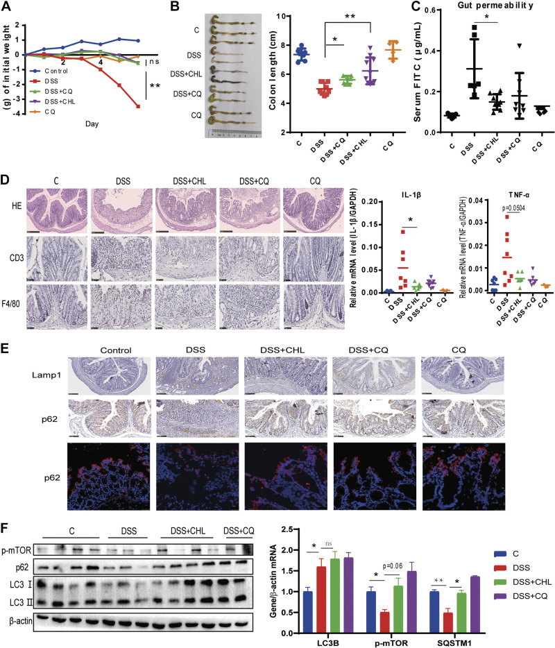
Figure 4.
Suppression of autophagy by chloroquine attenuates DSS-induced acute colitis in mice. Mice were treated with 3% DSS for 6 days. For several groups, CHL (40 mg/kg) was gavage from day 1 to day 6. Chloroquine (60 mg/kg) was intraperitoneally injected from day 1 to day 6. Saline as vehicle. (CQ group n = 4 animals, others n ≥ 6 animals). A and B: changes of body weight and colon length. C: gut permeability was determined by fluorescein isothiocyanate (FITC)-dextran presented in serum after oral gavage administration. D: representative images of colon tissues. Hematoxylin-eosin (H&E), CD3+ lymphocytes, and macrophages were measured via immunohistochemical staining. RT-qPCR analysis of mRNA levels of TNF-α and IL-1β. E: expression of p62/Sqstm1 and LAMP1 in the colon were determined by immunohistochemistry analysis and immunofluorescence. Scale bar = 100 μm. F: Western blot analysis of p-mTOR, LC3, and p62 of colon. The results shown are means ± SE. Statistical significance was determined by one-way ANOVA. *P < 0.05; **P < 0.01. DSS, dextran sulfate sodium.

