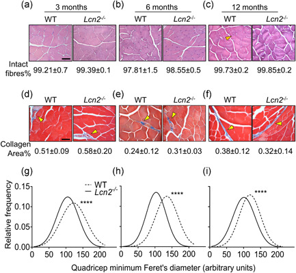
Figure 4.

Histological analysis of WT andLcn2 −/− mice quadriceps. Quadriceps from WT and Lcn2 −/− mice were explanted, fixed, paraffin‐embedded and sectioned at 5 μm. (a–c) Haematoxylin‐eosin staining performed on histological sections from (a) 3‐, (b) 6‐, and (c) 12‐month‐old mice to quantify the % of intact fibres. (d–f) Masson's trichrome staining to evaluate the % collagen area on histological sections from (d) 3‐, (e) 6‐ and (f) 12‐month‐old mice. (g–i) After haematoxylin‐eosin staining, minimum Feret's diameter from quadricep fibres was assessed via software (NIH ImageJ, version 1.50i.) in 3‐, (h) 6‐, and (i) 12‐month‐old mice. Gaussian curves were interpolated to better represent the fibre size distributions. (a–f) N = 3–5 mice per group; (g–i) N > 1000 fibres per group, arising from N = 3–5 mice per group. Scale bar = 50 μm; orange arrowhead: example of non‐intact (centrally nucleated) fibre; yellow arrowhead: collagen area stained in blue. Student's t test. ****p < .0001 versus WT. WT, wild‐type
