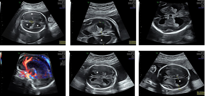
FIGURE 2.

Brain prenatal US imaging of patient 1 at 22 wGA, when hydrocephalus internus was apparent. The enlarged ventricles are highlighted by asterisks. The cerebellar size was described as being at the lower end of normal range, corpus callosum was normal (as shown by the Doppler US scan in the lower left corner)
