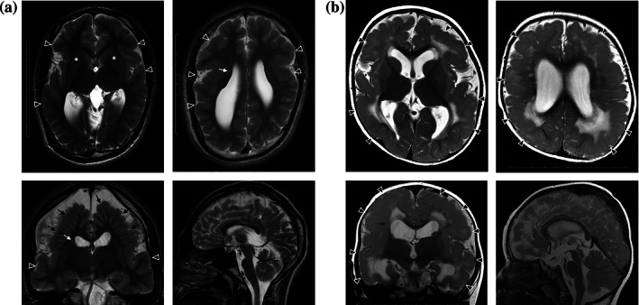
FIGURE 5.

Axial, coronal and sagittal T2‐weighted imaging (T2WI) of Patients 5 and 6 brain MRI. (a) Patient 5 brain MRI at 18 years of age showing dilatation of the lateral ventricles, perisylvian polymicrogyria (arrowheads), heterotopia (white arrows), radially‐oriented gyri (black arrows), and hypoplastic pyramidal tracts (asterisks). (b) Patient 6 brain MRI at 12 months of age, showing dilatation of the lateral ventricles, polymicrogyria (arrowheads) and high intensity signals in the deep white matter on T2WI
