
Figure 1.
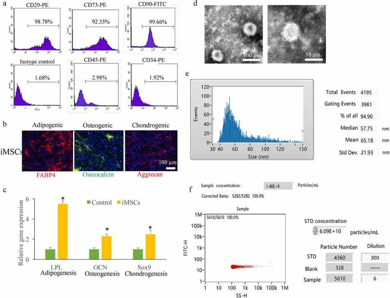
Identification iMSCs and isolated Exo. (a) The MSCs markers including CD29, CD90, CD73, CD45, and CD34 were measured. (b) The ability of iMSCs differentiating into chondrocytes, osteocytes, and adipocytes through staining aggrecan, osteocalcin, and FABP4. (c) OCN, Sox9, and LPL expression were measured. (d) The isolated exosomes were measured using transfer electron microscopy. (e) The average size of exosomes was measured by size distribution. (f) The exosomes concentrations were detected. * P < 0.05 compared with the group control. These results were obtained from at least three independent experiments. Sex-determining region Y-box 9 (Sox9); Osteocalcin (OCN); Lipoprotein lipase (LPL).