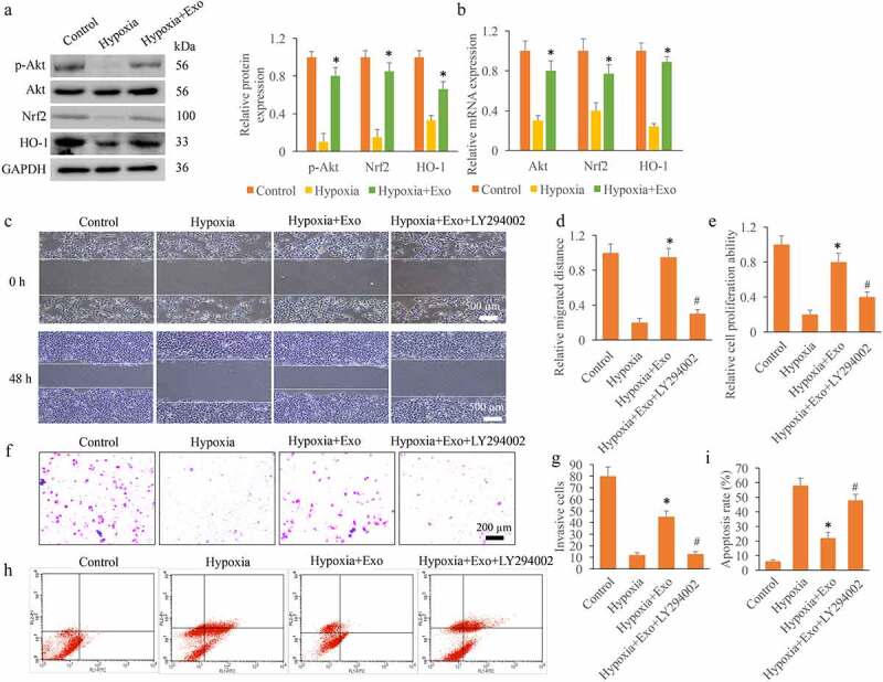
Figure 2.

Exo promoted cell viability through activating Akt/Nrf2/HO-1 signaling pathway in vitro. (a) The protein expression levels of p-Akt, Nrf2, and HO-1 were measured. (b) The mRNA expression levels of p-Akt, Nrf2, and HO-1 were measured. (c) The cell migration was measured with wound healing assay. (d) The cell migration was analyzed. (e) The cell proliferation was detected using CCK8 method. (f) The cell invasion was measured with Transwell method. (g) The cell invasion was analyzed. (h) The cell apoptosis was detected with flow cytometry. (i) The cell apoptosis was analyzed. * P < 0.05 compared with group hypoxia. # P < 0.05 compared with the group hypoxia+Exo. These results were obtained from at least three independent experiments.
