
Figure 2.
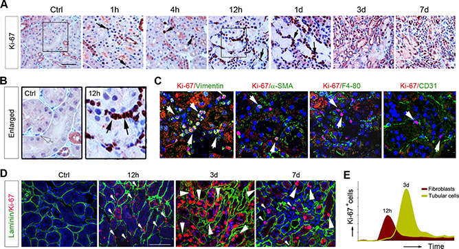
Fibroblast proliferation precedes tubular repair and regeneration after AKI. A, B) Immunohistochemical staining for Ki‐67 showed that fibroblast proliferation is an early event preceding tubular regeneration. A) Representative micrographs of renal Ki‐67 staining at different time points after IRI are shown. Scale bar, 50 μm. B) Enlarged boxes are presented. Arrows indicate Ki‐67‐positive cells in renal interstitium. C) Double immunostaining showed renal Ki‐67 and cell type‐specific markers, such as vimentin, α‐SMA, F4/80, and CD31, at 12 h after IRI. White arrows indicate Ki‐67‐positive cells. D) Coimmunostaining with antibodies against Ki‐67 and laminin demonstrated tissue compartment‐specific cell proliferation at different time points after IRI. Small arrowheads indicate interstitial cells, whereas large arrowheads indicate tubular epithelial cells. E) Graphic presentation showed different dynamics of fibroblast and tubular cell proliferation after AKI. Ctrl, control.