Figure 3.

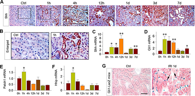
Tubule‐derived Shh triggers fibroblast activation in AKI. A, B) Shh was induced specifically in renal tubules as early as 1 h after IRI. A) Kidney sections at different time points after IRI were immunostained with anti‐Shh antibody. Scale bar, 50 μm. B) Boxed areas were enlarged and presented. Arrow indicates positive staining. C‐F) Shh signaling was rapidly activated in the kidney after AKI. Renal expression of Shh (C), Gli1 (D), Patch1 (E), and Hhip (F) mRNA at different time points after IRI was assessed by real‐time qPCR. **P < 0.01, *P < 0.05 vs. sham‐treated controls (n =3‐4). G) Shh targets interstitial fibroblasts in AKI. The Gli1‐LacZ reporter mice were subjected to IRI. At 1 d after IRI, kidney sections were subjected to X‐gal staining. Arrows indicate X‐gal‐positive interstitial cells in renal interstitium. Scale bar, 50 μm. Ctrl, control.
