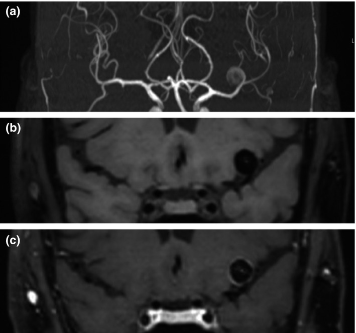
FIGURE 1.

Example of aneurysm wall enhancement in a patient with a left middle cerebral artery aneurysm. All images are coronal projections and were made during the same scanning session. (a) Coronal maximum intensity projection of 3D time‐of‐flight MRA. High spatial resolution, fat‐saturated 3D T1 SPACE black blood MRI before (b) and after (c) administration of gadolinium, showing circumferential aneurysm wall enhancement
