
FIGURE 1.
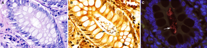
Photomicrograph of the colonic mucosa of a healthy dog. The bacteria within the crypts of healthy dogs are inconspicuous on routine hematoxylin and eosin stain (A). The Steiner silver stain (B) highlights abundant bacteria (arrow) within the crypts. Fluorescence in situ hybridization with EUB338 probe targeting all bacteria in the crypts. Labeled bacteria appear red (arrow). The autofluorescence of the intestinal mucosa appears green. DAPI (4′,6‐diamidino‐2‐phenylindole)‐stained nuclei of colonic mucosa appear blue. ×60 objective. Courtesy of Dr Paula Giaretta, DACVP, Universidade Federal de Minas Gerais, Brazil 27