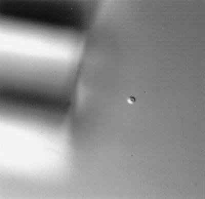
FIG. 1.
Differential interference contrast microscopy of a single C. parvum oocyst during micromanipulation. Magnification, ×400. FITC-labeled oocysts were located using epifluorescence microscopy and isolated with 100× phase microscopy using a manually pulled glass micropipette (≈20-μm-inner-diameter tip opening) together with a micropump stabilized by a micromanipulator.

