FIGURE 5.

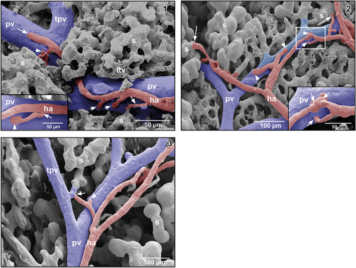
(1) Hepatic arteriole (ha) running along a portal venule. Note that the arteriole first gives off a branch to a sinusoid forming an arteriolar‐sinusoidal anastomosis (ASA; small arrow), then two branches to the fellow portal venule forming arteriolar‐portal (venous) anastomoses (APAs; arrowheads) to finally anastomose with the fellow portal venule (large arrow). Inset Short arteriolar‐portal anastomosis (APA). Note the widened inlet portion (arrowhead) and the narrowing of the hepatic arteriole at its origin from the parent hepatic arteriole (arrow). (2) Arteriolar‐portal (APAs) and ASAs. Note spacing and trumpet‐like shape of the arteriolar inlet portions in APAs (arrowheads) and ASAs (arrows). Inset Enlargment of the framed area displaying three closely spaced APAs (arrows). (3) Hepatic arteriole joining a tubular inlet portion of a sinusoid (short arrow). The long arrow points at an arteriole that in this angle of view seems to join the portal venule, but does not do so. This was confirmed by changing the tilting angle during SEM work
